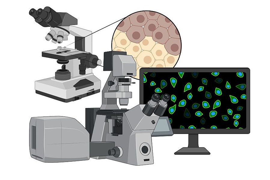
The Microscopy Facility offers a comprehensive range of state-of-the-art light microscopes and software solutions for image analysis. Experienced staff provide support in the planning, acquisition and evaluation of imaging analysis methods.
Our services:
- Preliminary discussions and advice on imaging experiments
- Support in planning and sample preparation
- Training on light microscopy systems and analysis softwares
- Assistance with microscopy
- Image data processing and analysis
We are also involved in the maintenance and upgrade of existing systems, as well as in the acquisition process of new systems. This includes organizing demos, subsequent testing of systems and determining the required components of complex imaging systems.
Our methods:
- Bright field microscopy
- Wide field fluorescence microscopy
- Confocal microscopy
- Live cell time lapse microscopy
- Spinning disc confocal microscopy
- Digitization of histological samples
- Image-based cytometry / high content screening
- Automated imaging
Our systems:
- Various wide field fluorescence microscopes, upright + inverted, incl. cameras
- Visitron Live cell phase contrast & wide field fluorescence microscope incl. incubation, 2D FRAP
- Zeiss LSM700 laser scanning confocal microscope
- Olympus spinning disc confocal microscope with Yokogawa unit (50µm + SoRa disc), incubation, ScanR high content screening software
- Incucyte S3 + Wound maker Tool
- 3D Histech SCAN II bright field slide scanner
- 3D Histech MIDI fluorescence slide scanner
- Huygens Deconvolution Software
- Definiens Tissue Studio
- etc.
To allow us to provide best possible support and imaging quality, please involve us in planning of your experimental setup and contact us timely.
Email of the CF microscopy & imaging: imaging-CCR@meduniwien.ac.at

Dominik Kirchhofer, BSc

