
Histology is an important pillar in the diagnosis of pathological tissue changes in both medicine and research. For this reason, it is an important tool, especially in cancer research, for preparing tissue and making fine-tissue structures visible microscopically.
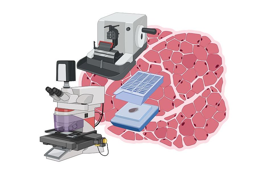
Our main tasks include the pre-treatment of the tissue in the tissue-embedding machine and the embedding of the fixed tissue in paraffin, which subsequently enables the histological sectioning on the microtome. In this process, tissue sections in the micrometer range are generated so that individual cell layers of the tissue can be made visible. In addition, a histological staining method, such as hematoxylin-eosin staining, allows visualisation of cellular structures of the cell layers at the microscopic level. Alternatively, immunohistochemistry offers the possibility to specifically detect and visualise antigens in the tissue section.

Martin Filipits, Assoc. Prof. Univ.-Doz. Mag. Dr.
